- Afrikaans
- Albanees
- Amharies
- Arabies
- Armeens
- Azerbeidjans
- Baskies
- Wit-Russies
- Bengaals
- Bosnies
- Bulgaars
- Katalaans
- Cebuano
- China
- Korsikaans
- Kroaties
- Tsjeggies
- Deens
- Nederlands
- Engels
- Esperanto
- Estnies
- Fins
- Frans
- Fries
- Galisies
- Georgies
- Duits
- Grieks
- Gujarati
- Haïtiaanse Kreools
- Hausa
- hawaiian
- Hebreeus
- Nee
- Miao
- Hongaars
- Yslands
- igbo
- Indonesies
- iers
- Italiaans
- Japannese
- Javaans
- Kannada
- kazaks
- Khmer
- Rwandese
- Koreaans
- Koerdies
- Kirgisies
- Arbeid
- Latyn
- Letties
- Litaus
- Luxemburgs
- Masedonies
- Malgassies
- Maleis
- Malabaars
- Maltees
- Maori
- Marathi
- Mongoolse
- Myanmar
- Nepalees
- Noors
- Noors
- Oksitaans
- Pasjto
- Persies
- Pools
- Portugees
- Punjabi
- Roemeens
- Russies
- Samoaans
- Skotse Gaelies
- Serwies
- Engels
- Shona
- Sindhi
- Sinhala
- Slowaaks
- Sloweens
- Somalies
- Spaans
- Soendanees
- Swahili
- Sweeds
- Tagalog
- Tadjieks
- Tamil
- Tataars
- Telugu
- Thai
- Turks
- Turkmeens
- Oekraïens
- Oerdoe
- Uighur
- Oezbeeks
- Viëtnamees
- Wallies
- Help
- Jiddisj
- Yoruba
- Zoeloe
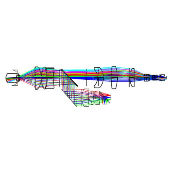
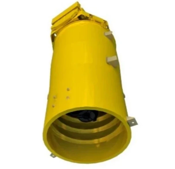
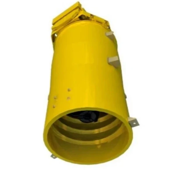
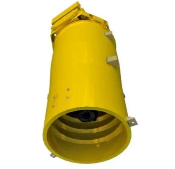
Fundus Beelder
Produkte Besonderhede

Fundus Imager Hoof Tegniese Aanwysers
|
Werksafstand |
> 24 mm |
|
Beeldmodus |
Naby-infrarooi waarneming, fokus, fotografie met sigbare lig |
|
Leerling Grootte |
Verwydingsvry 3mm |
|
Retinale resolusie |
Central field of view better than 7 μ m, edge field of view better than 15 μ m |
|
Brekingskompensasiereeks |
-20D~+20D |
|
Gesigveld |
45° |
|
Beeldspektrum |
Sigbare lig: RGB-kleur (450-700) Naby infrarooi: 780nm Bevestigingslig: 650nm |
|
MTF |
Greater than 0.2@resolution corresponding image side line pairs |
The Fundus Imager is a cutting-edge diagnostic tool designed to capture high-resolution images of the retina, aiding in the early detection and monitoring of various eye conditions such as diabetic retinopathy, glaucoma, macular degeneration, and retinal diseases. It utilizes non-mydriatic imaging technology, enabling the capture of detailed fundus images without the need for pupil dilation, ensuring a comfortable and efficient examination for patients. The imager features a high-definition camera, advanced optical components, and LED lighting, which provide clear and accurate images of the retina, optic disc, and macula. With its automated alignment and user-friendly interface, it allows for easy operation, even by less experienced staff, and can produce instantaneous results, making it an invaluable tool in clinical practice. The device is also compatible with various imaging systems and electronic health record (EHR) platforms, facilitating smooth integration into modern healthcare workflows.
The key advantages of the Fundus Imager include its ability to produce high-quality, detailed retinal images without the discomfort of pupil dilation, allowing for faster patient throughput and enhanced patient satisfaction. Its non-invasive nature and quick image capture make it ideal for routine eye exams and large-scale screenings. Additionally, its compact design and ease of use ensure that it is accessible for both small clinics and large hospitals. The real-time imaging and automated features significantly improve diagnostic accuracy, enabling healthcare providers to make informed decisions faster, ultimately contributing to early detection and effective management of ocular diseases.
